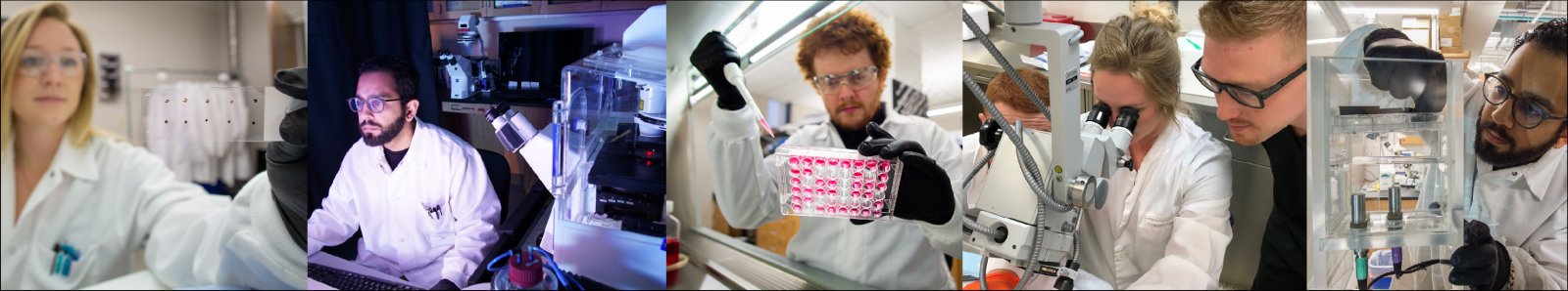
Overview
Research in the Translational Mechanobiology Lab (TML) employs techniques in biomechanics, cell and molecular biology, histology, imaging, regenerative medicine, and tissue engineering to explore the influence of biomechanical (and biochemical) stimuli in modulating normal and pathological cell behaviors. The goal is to translate an understanding of mechanobiological mechanisms of disease to create new therapies, develop prognostic indicators of disease progression, and improve the design of implantable medical devices. The primary applications of the work are in cardiovascular medicine, ophthalmology, and wound healing.
Atherosclerosis
Atherosclerosis is a chronic inflammatory disease that manifests as plaques composed of a lipid-rich necrotic core and immune cells within the intima of the artery wall. Plaque rupture exposes the highly thrombogenic necrotic core to the bloodstream, leading to platelet activation, thrombosis, and artery occlusion. This event is the most common trigger of heart attack and stroke, which are leading causes of morbidity and mortality in the United States. An interesting feature of atherosclerosis is that plaques develop in regions of arteries experiencing disturbed blood flow. This observation suggests that specific biomechanical signatures play a principal role in atherogenesis by promoting a dysfunctional endothelium—the cell type that sits at the interface between the bloodstream and vessel wall.

Characterizing mechanotransduction signaling pathways in endothelial cells under normal and atherogenic arterial mechanical environments
The precise environmental cues that promote advanced plaque development, particularly those vulnerable to rupture, remain unknown. Our laboratory explores how different biomechanical signatures (stemming from disturbed vessel wall stretch and blood flow) activate mechanotransduction signaling in dysfunctional endothelial cells to promote the development of advanced plaques. The goal is to identify novel pharmaceutical targets of patho-mechanotransduction in atherosclerosis.
Engineering mechanotherapies as a regenerative medicine approach to treat endothelial cell dysfunction in advanced atherosclerotic plaques
Assessing the efficacy of nanoparticles as both a diagnostic and therapeutic for advanced plaques

Developing prognostic indicators of plaque progression
Several studies, including from our group, have quantified a relationship between local blood flow disturbances and the development of specific plaque types in both patients and experimental animal models. Our laboratory is working to synthesize these data within a computational framework to predict plaque progression. This framework includes the development of novel mechanical biomarkers and the use of techniques in statistical pattern recognition and machine learning.Cataract Surgery
Cataract surgery is the most common corrective procedure in the aged population and post-surgical capsule fibrosis is the most common complication. During surgery, the cloudy lens fibers are removed through a permanent hole placed in the lens capsule and replaced with an artificial intraocular lens (IOL). This procedure permanently alters the biomechanical environment of the lens capsule, which promotes the fibrotic response that is mediated by the inhabiting lens epithelial cells. The severity of capsule fibrosis depends on IOL design and current IOLs lack the ability to accommodate (i.e., change focus from distant to near objects).
